PUNE: A woman got back her sight eight months after a Pune-based eye surgeon harvested her outer
retinal tissue and grafted it to plug a hole in her eye, and the recurrent retinal detachment.
It is probably the first such surgery in Pune. Glaucoma robbed the patient, a 47-year-old woman from Urali Kanchan, some 32 km from Pune, of her left eye, and an injury from the sharp edge of a dining table tore the retina of her right eye.
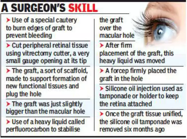
She underwent vitrectomy but it failed to fix the trauma-led hole in the central part of her retina
(macula). Due to recurrence of the retinal detachment, she had loss of vision. The retina is a nervous
tissue and cannot be replaced with an artificial one.
The vision restoration suggests that the retinal graft placed inside the macular hole has led to the formation of new retinal tissues and repaired the hole in the fovea , the centre of the macula.
“Functional recovery after surgery is often gradual. Over the last eight months, the near-blind woman who only had light perception has achieved 6/60 vision as against the normal 20/20 vision. That means she can see or perceive objects within 15 to 20 feet and has met the criterion of a ‘walking vision’ which is a remarkable improvement,” eye surgeon Aditya Kelkar, director of the National Institute of Ophthalmology, told TOI.
Kelkar said it is a first in Pune using an autologous graft although a few surgeons in India have attempted it with some success in the past.
“Further studies are certainly needed to investigate if autologous retinal graft could not only serve as a scaffold but also help retain some function and promote the reparation of the retinal layers in other cases of retinal tears or holes,” Kelkar said.
The woman said the vitrectomy two years ago temporarily restored some amount of vision but it didn’t last long. “I was almost blind a year after the surgery and was dependent on others,” she said.
The second surgery eight months ago required out-of-the-box thinking. Kelkar decided to carry it out in two stages.
” Laser treatment was not suitable since the detachment was recurrent and the hole was located in the centre of retina. The laser could have led to scarring defeating the entire purpose,” he said.
This surgery was done using the microscope called artevo, a digital microscope that allowed 3D visualization of retina on a big screen. The intra operative Optical Coherence Tomography made the surgery helped the surgeon see the exact location of the placement of graft tissue and attachment of retina.